Subtalar Joint Fusion (Arthrodesis)
Examples: Case 1 | Case 2
Arthritis is when the cartilage is damaged or worn away and no longer provides the cushion to allow the smooth motion between the bones. Cartilage is unique in that there is no blood flow that provides nutrition, so that when injured it does not have the ability to repair itself very well, unlike bone. This inability to repair itself is why scientists and surgeons have a very difficult time in trying to restore large cartilage defects in the setting of arthritis. When non-surgical options such as injection, braces, and activity modification have not provided sufficient relief, surgery can be considered. Patients with subtalar arthritis have pain that is noted on the side of the ankle/hindfoot and have more difficulty with uneven ground, grass, or gravel. There is no replacement option for this joint and the most effective method to surgically treat arthritis of this joint is a fusion.
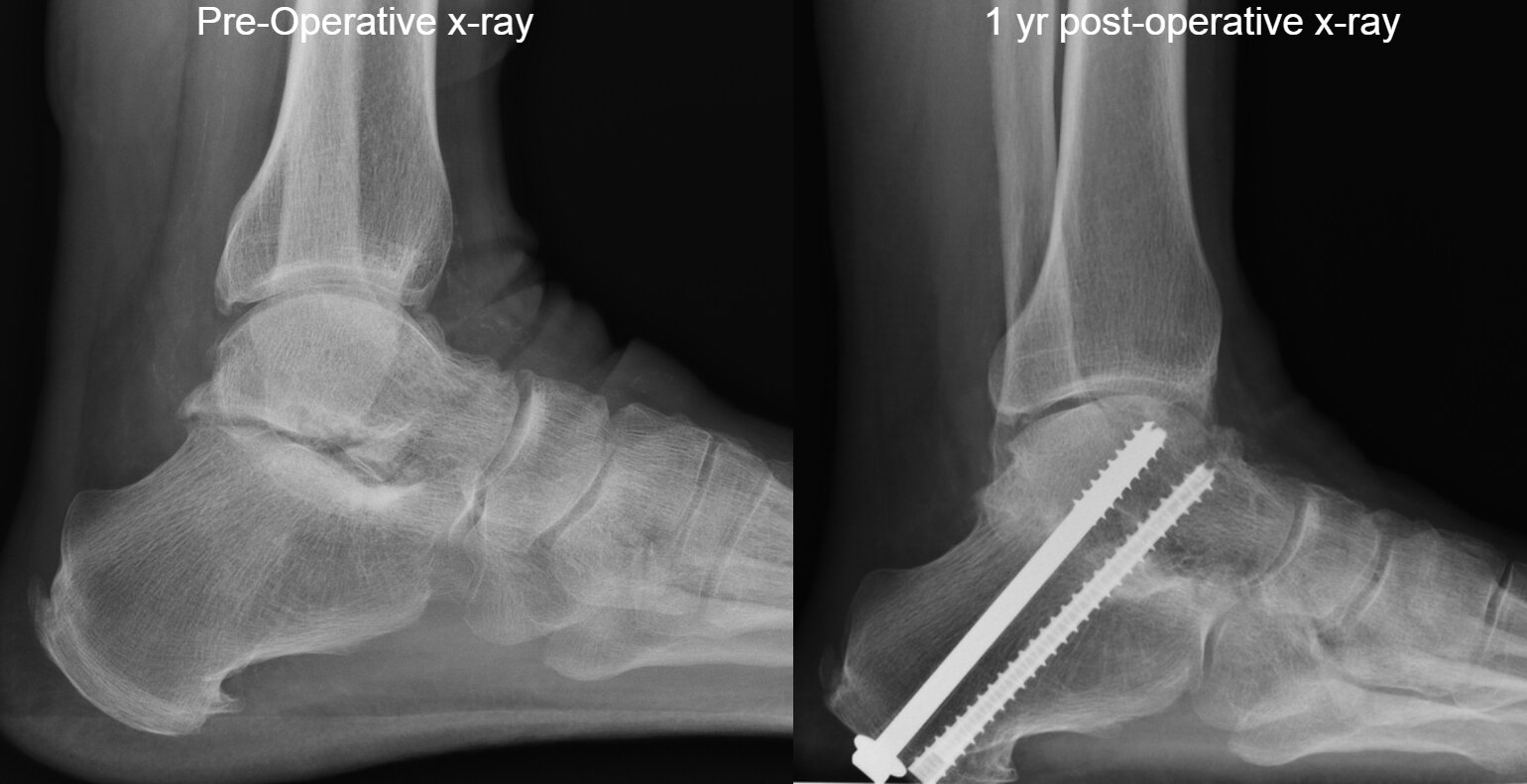
A subtalar fusion is most commonly an outpatient operation and “glues” the two bones together - the talus and calcaneus. The goal of the fusion is to make the body connect these two bones that were previously separated by cartilage with solid bone. Normally, two bones are separated by cartilage also known as a joint. Arthritis causes normal cartilage to be worn out and causes painful bone on bone motion in the joint. With fusion, two bones are fused into one bone in order to eliminate any motion/joint as the two bones become one larger bone, resulting in pain relief. However, the major downside of a fusion is the loss of motion of the hindfoot. The entire foot is not fused, so there remains a minimal amount of the side to side motion of the entire foot. In patients who have a very stiff subtalar joint, the loss of function is minimal for them as they have not had motion in their hindfoot/subtalar joint for years. Patients who have a lot of subtalar/hindfoot motion (side to side ) will notice the stiffness after surgery following a fusion.
We use an open approach to ensure the joint is positioned appropriately and the deformity is corrected so that the foot is in a neutral position. The remaining cartilage is removed and bone is “prepared” to get the fusion to heal. Preparation of the bones means that small drill holes and microfractures are made to stimulate bone healing and bone graft is added to maximize the chance of a fusion. Multiple screws are commonly used to hold the bones in the right position and pressed together. Over time the bones will heal - and this is the successful fusion that we hope for. If there is significant bone loss that may occur from a calcaneus fracture, a larger bone graft that is structural to restore the height and anatomy of the foot may be required. In this particular case, the period of no pressure on the foot (non-weightbearing) may be increased from 6 weeks to 3 months given the complexity of healing required. The screws are not actually holding the bone together in the long-term, it is the new bone that has formed between the talus and calcaneus and this is why it can last a lifetime once healed.
Following surgery, a successful fusion occurs in the majority of patients resulting in relief of pain. Patients are able to walk, bike, swim without significant difficulty in most cases. Shoewear has some limitations, but any comfortable athletic shoe and occasionally fashionable shoes for short time periods can be used. Over time, average of 20 years, patients may develop arthritis in the joints surrounding the fusion and some pain can result from this arthritis. If the fusion does not occur and the two bones do not grow together, that can occur despite an appropriately done operation, there may be persistent swelling and pain that requires a revision of the surgery.
Outcomes of Subtalar joint fusion based on published literature:
Returned to level of activities prior to symptoms: 84% (Reference)
No improvement in symptoms after surgery - 16% (Reference)
Union rate after primary subtalar fusion - 86% (Reference), 98% (Reference)
Union rate after Revision subtalar fusion - 71% (Reference)
Nonunion - 2% (Reference), 6.3% (Reference), 7% (Reference). 21.5% (Reference)
Prominent hardware requiring removal of hardware - 8.4% (Reference), 13% (Reference), 27% (Reference)
Adjacent joint arthritis (Talonavicular Joint, Tibiotalar joint) - 13.5% (Reference)
Superficial Wound Infection - 6.3% (Reference)
Deep venous thrombosis - 2.1% (Reference)
Related Topics:
Our Published Literature Related to Subtalar Joint Fusion
Subtalar Arthrodesis. Reconstructive Foot & Ankle Surgery: Management Of Complications. (2019) - Book Chapter
Triple Arthrodesis. Reconstructive Foot & Ankle Surgery: Management Of Complications. (2019) - Book Chapter
Tibiotalocalcaneal & Pantalar Arthrodesis. Reconstructive Foot & Ankle Surgery: Management Of Complications. (2019) - Book Chapter
Tarsal Coalition. Reconstructive Foot & Ankle Surgery: Management Of Complications. (2019) - Book Chapter
Disorders Of The Foot & Ankle. Miller’s Review Of Orthopaedics. (2019) - Book Chapter
Overview Of The Ankle & Foot. Essential Orthopaedics. (2019) - Book Chapter
Imaging Of The Foot & Ankle. DeLee & Drez’s Orthopaedic Sports Medicine. (2018) - Book Chapter
Hindfoot. Passport For The Orthopedic Boards & FRCS Examination. (2015) - Book Chapter
Tarsal Coalition. Presentation, Imaging & Treatment Of Common Musculoskeletal Conditions. (2011) - Book Chapter
Malunion & Nonunion In Foot & Ankle Surgery. Foot & Ankle Specialist. (2010)
Percutaneous Screw Configuration Versus Perimeter Plating Of Calcaneus Fractures: A Cadaver Study. Foot & Ankle International. (2008)
Ankle & Hindfoot Reconstruction: What Is New In Ankle Arthroplasty, Allograft, & Fusion. Current Opinion In Orthopaedics. (2004)
Hindfoot Arthrodesis For The Adult Acquired Flat Foot. Foot & Ankle Clinics. (2003)