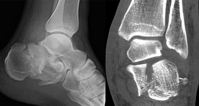
The heel bone/calcaneus is the first bone that contacts the ground when walking. A significant amount of weight is transferred to this bone during heel strike (when the heel contacts the ground). The joint above this bone - called the subtalar joint is very important for the side to side motion of the foot. The bone also has another joint called the calcaneo-cuboid joint that also aids in the side to side motion. The tendons that help to control the motion of the foot for side to side motion and for pulling the toes down are adjacent to the bone. The bone itself has a very thin shell covering spongy type bone. In cases of a fracture, it can range from a simple fracture that has not shifted in mild cases, to severe cases where the bone is multiple fragments, severe deformity with the heel getting wider, shorter, and tilted in. The cartilage of the subtalar joint and the calcaneocuboid joint can be severely damaged in these cases. Cartilage damage is not repairable and this is the primary reason why arthritis occurs from these fractures, despite the bones being put back together.
Patients who suffer this injury have been through a high energy trauma, commonly a fall from a height, such as a ladder or even a roof, or a car accident. Associated fractures with the spine can occur from such severe injuries. An examination is done to evaluate the status the skin and to determine if any other injuries have occurred besides the calcaneus fracture. Xrays are reviewed and additional imaging with a CT scan is needed to determine the alignment of the bones, the severity of the fracture, and to determine what surgical intervention may be appropriate.
Treatment of calcaneus fractures does not always require surgery. Timing is critical as well, the sooner we can surgically address the fracture, the more likely smaller incisions can be used. Our office will find time for you if you call with this injury and add fractures on within 24 hours in most cases. Surgical intervention is dependent upon the severity of the fracture, medical condition of the patient, activity demands, among other considerations. The primary difficulty with this fracture is the soft tissue/skin and surgery does carry a higher than average risk of wound complications when comparing to most other fractures. Traditionally, a large incision is made along the side of the foot to expose the broken bone and put it back together with plates and screws. Although this method does allow for good exposure of the fracture, there is a significant risk of wound complication and therefore we do not use this approach. Recent literature has shown that a large open approach does not offer significant benefit compared to non-surgical treatment. We employ modern techniques using smaller incisions or in some cases - percutaneous (small 1 cm incisions) to put the fracture back into alignment and hold the position with screws and specially made plates for small incisions. The goal of treatment is to realign the shape of the foot the best that is possible, minimize the risk of arthritis, and to minimize the risk of infection. Despite all treatment efforts, the risk of arthritis is still high with surgery, but if the foot is realigned, the risk is lower and surgery to fuse the joint to treat the arthritis is better for the patient if the anatomy of foot is restored. In all cases, the decision to pursue surgery or non-surgical treatment is up to the patient and we do our best to provide you with the best information possible to make this decision.